


Therapeutic area
Oncology
Thank you for your interest!
9M+
journeys for patients with cancer, including pediatric populations
3B+
clinical notes, including pathology reports, radiology reports, and oncologist progress notes
94M+
imaging studies
Study all common and rare cancers with unparalleled breadth and depth
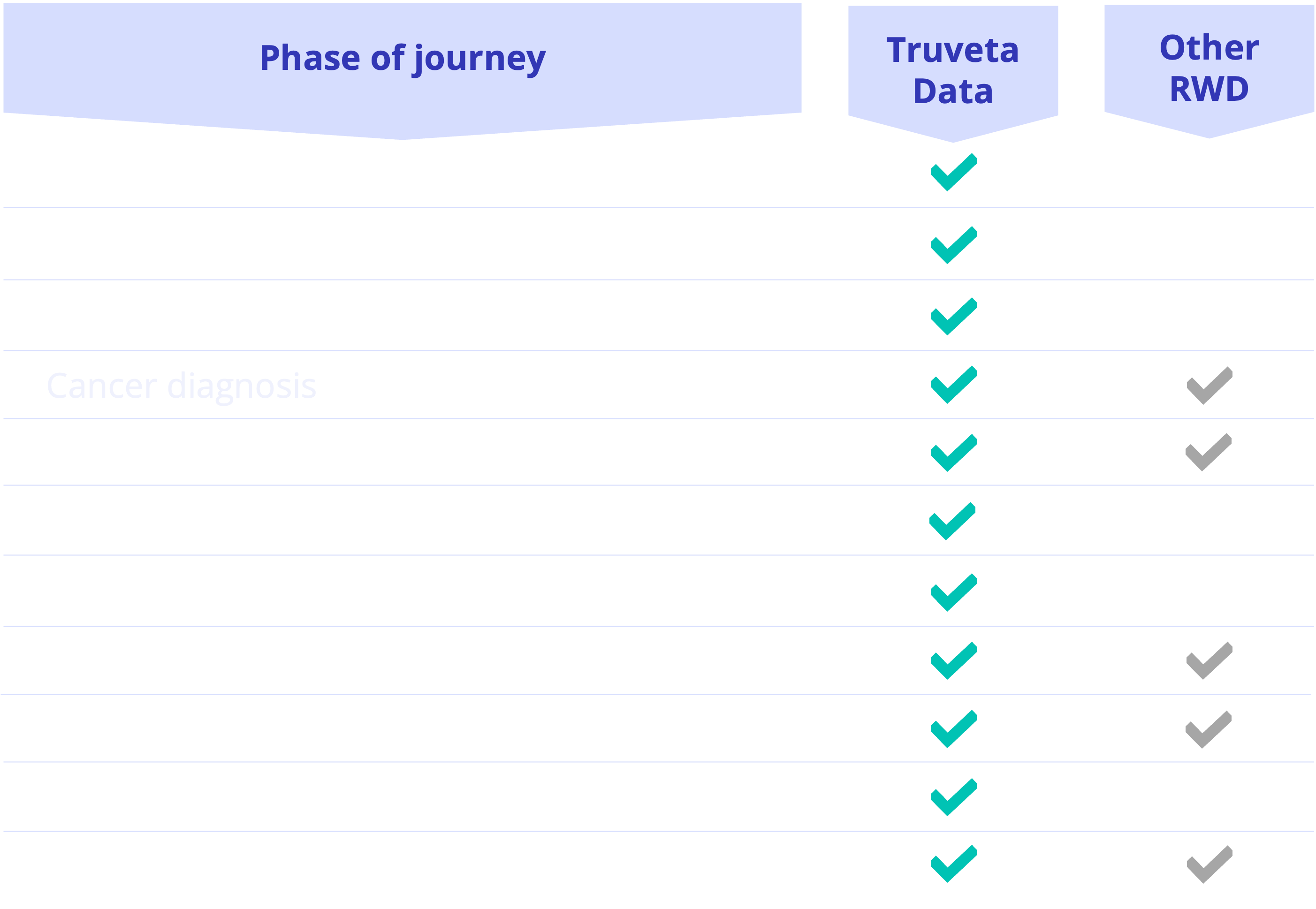
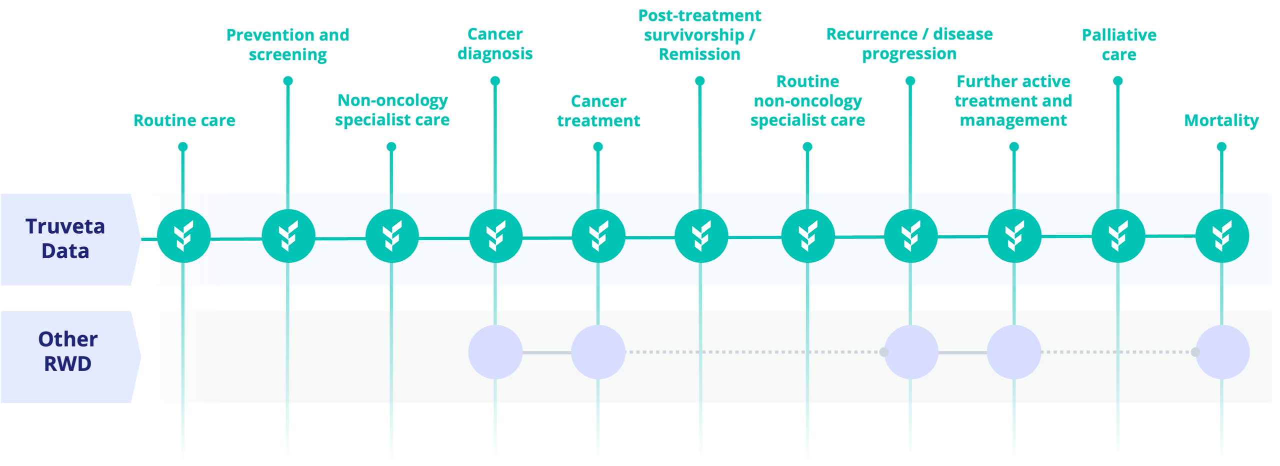
Explore the complete patient journey from symptoms and diagnosis through treatment, progression, recurrence, survivorship, and long-term outcomes.
Disease characterization
- Stage, grade, and histology
- Biomarkers and disease characteristics
- Radiographic images
- Pathology images
Treatment and disease course
- Therapies and procedures
- Treatment response
- Toxicity and side effects
- Discontinuation and reason
- Disease progression and recurrence
- Mortality
Patient context
- Comorbidities
- Social determinants of health
- Performance status
- Race/ethnicity
- Family history
- Risk factors

Disease characterization

Treatment and disease course

Patient context
Disease characterization

Treatment and disease course
Patient context
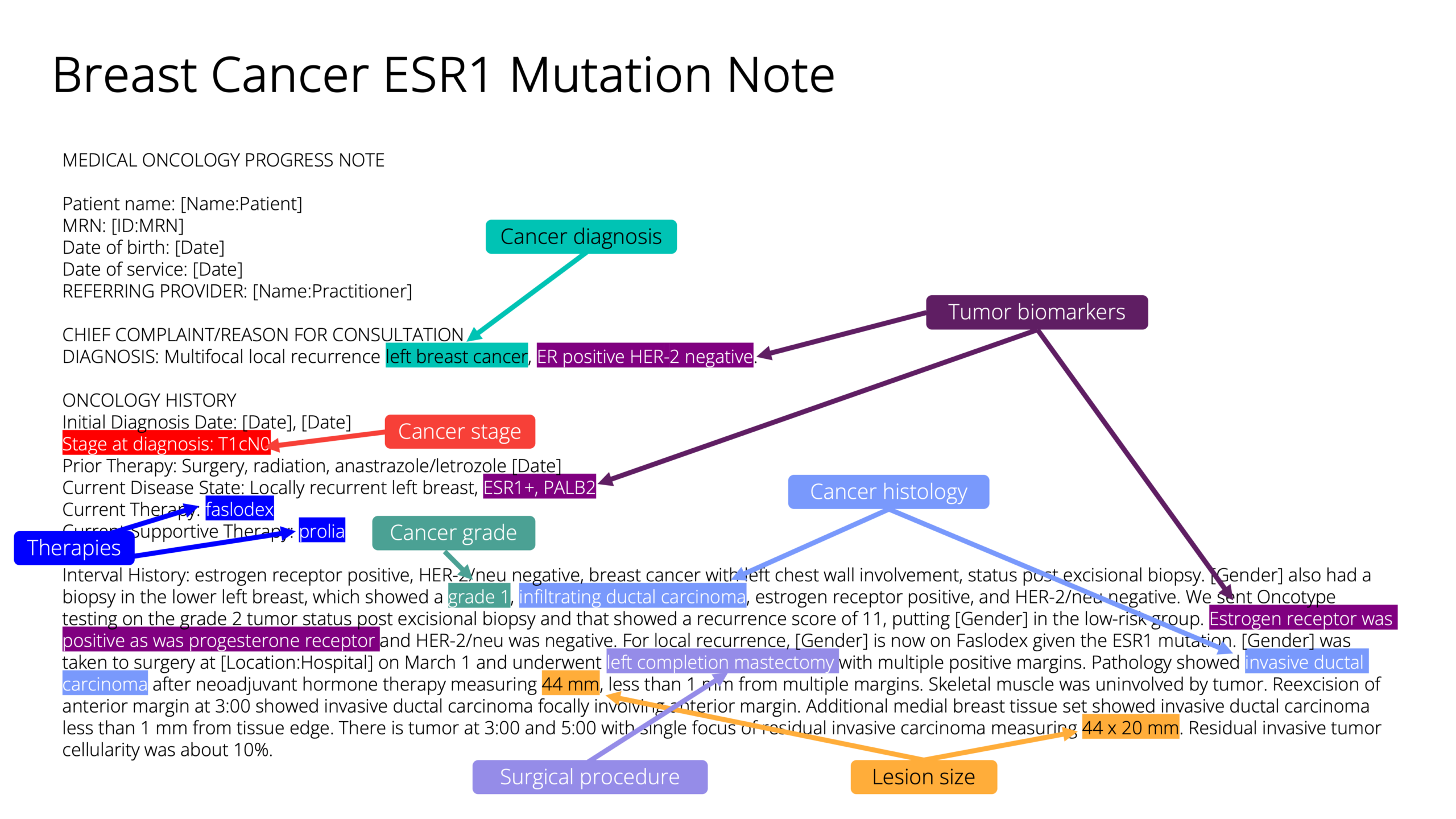
Unlock the complexity of oncology with notes
Integrated with structured EHR data and longitudinally integrated with notes and images, these validated variables enable richer patient characterization and more accurate longitudinal oncology research.
Visualize cancer care and train AI with integrated images
Connect rich clinical context with imaging and waveform data, searchable by modality and protocol—including DICOM images with pixel and metadata—across the complete patient journey. Enable deeper analysis of cardiac function, rhythm, procedures, and care events in a single, longitudinal view.
Accelerate insights to real-world impact
AI training
Clinical trials
Accelerate oncology product development with real-time tracking of trial-eligible patients and sites or build confirmatory evidence
Therapy adoption
Optimize commercial performance with the ability to monitor treatment utilization and variation in real time
Safety
Save on post-market requirements by detecting complications and adverse earlier from clinically deep, longitudinal patient journeys
Outcomes research
Generate differentiated evidence and evaluate clinical and economic outcomes with more depth
Sample of cancers represented in Truveta Data
Breast
391K
Total population
Prostate
862K
Total population
Glioblastoma
20K
Total population
Lung
652K
Total population
Gastric
70K
Total population
Non-Hodgkin lymphoma
391K
Total population
Cervical
122K
Total population
Thyroid
191K
Total population
Colorectal
459K
Total population
Oropharyngeal
170K
Total population
Liver
123K
Total population
Melanoma
432K
Total population
A
- Acinar cell carcinoma
- Acute lymphoblastic leukemia (ALL)
- Acute myeloid leukemia (AML)
- Adenocarcinoma
- Anal cancer
- Anaplastic thyroid cancer
- Angiosarcoma of the liver
B
- Basal cell carcinoma
- Bladder cancer
- Bone sarcomas – Chondrosarcoma
- Bone sarcomas – Ewing sarcoma
- Bone sarcomas – Osteosarcoma
C
- Cervical cancer
- Cholangiocarcinoma (bile duct cancer)
- Chromophobe renal cell carcinoma
- Chronic lymphocytic leukemia (CLL)
- Chronic myeloid leukemia (CML)
- Clear cell renal cell carcinoma
- Collecting duct carcinoma
- Cutaneous T-cell lymphoma
D
- Ductal carcinoma in situ (DCIS)
E
- Epithelial ovarian cancers – Clear cell carcinoma
- Epithelial ovarian cancers – Endometrioid carcinoma
- Epithelial ovarian cancers – Mucinous carcinoma
- Epithelial ovarian cancers – Serous carcinoma (high-grade and low-grade)
F
- Familial adenomatous polyposis (FAP)
- Fibrolamellar carcinoma
- Follicular thyroid cancer
G
- Gallbladder cancer
- Germ cell tumors
- Gliomas – Astrocytoma
- Gliomas – Glioblastoma
- Gliomas – Oligodendroglioma
H
- Hairy cell leukemia
- Hepatoblastoma
- Hepatocellular carcinoma
- HER2-positive breast cancer
- Hodgkin lymphoma – Lymphocyte-depleted
- Hodgkin lymphoma – Lymphocyte-rich
- Hodgkin lymphoma – Mixed cellularity
- Hodgkin lymphoma – Nodular sclerosis
- Hormone receptor-positive breast cancer
- Hypopharyngeal cancer
I
- IgA myeloma
- IgG myeloma
- Inflammatory breast cancer
- Invasive ductal carcinoma (IDC)
- Invasive lobular carcinoma (ILC)
L
- Laryngeal cancer
- Light chain myeloma
- Lynch syndrome
M
- Medullary carcinoma
- Medullary thyroid cancer
- Medulloblastoma
- Melanoma – Acral lentiginous
- Melanoma – Lentigo maligna
- Melanoma – Nodular
- Melanoma – Superficial spreading
- Meningioma
- Merkel cell carcinoma
- Mucinous adenocarcinoma
N
- Nasopharyngeal cancer
- Neuroendocrine tumors
- Non-Hodgkin lymphoma – Burkitt lymphoma
- Non-Hodgkin lymphoma – Diffuse large B-cell lymphoma
- Non-Hodgkin lymphoma – Follicular lymphoma
- Non-Hodgkin lymphoma – Mantle cell lymphoma
- Non-Hodgkin lymphoma – Marginal zone lymphoma
- Non-secretory myeloma
- Non-small cell lung cancer (NSCLC) – Adenocarcinoma
- Non-small cell lung cancer (NSCLC) – Large cell carcinoma
- Non-small cell lung cancer (NSCLC) – Squamous cell carcinoma
O
- Oncocytic carcinoma
- Oral cavity cancer
- Oropharyngeal cancer
P
- Paget’s disease of the breast
- Pancreatic ductal adenocarcinoma
- Pancreatic neuroendocrine tumors (PNETs) – Gastrinoma
- Pancreatic neuroendocrine tumors (PNETs) – Glucagonoma
- Pancreatic neuroendocrine tumors (PNETs) – Insulinoma
- Pancreatic neuroendocrine tumors (PNETs) – Non-functioning PNETs
- Papillary renal cell carcinoma
- Papillary thyroid cancer
- Pituitary adenomas
- Plasma cell leukemia
R
- Renal medullary carcinoma
- Retinoblastoma
S
- Salivary gland cancers – Acinic cell carcinoma
- Salivary gland cancers – Adenoid cystic carcinoma
- Salivary gland cancers – Mucoepidermoid carcinoma
- Schwannomas
- Sex cord-stromal tumors
- Signet ring cell carcinoma
- Small cell lung cancer (SCLC) – Combined small cell carcinoma
- Small cell lung cancer (SCLC) – Small cell carcinoma
- Smoldering multiple myeloma
- Soft tissue sarcomas – Angiosarcoma
- Soft tissue sarcomas – Leiomyosarcoma
- Soft tissue sarcomas – Liposarcoma
- Soft tissue sarcomas – Rhabdomyosarcoma
- Soft tissue sarcomas – Synovial sarcoma
- Squamous cell carcinoma
T
- Triple-negative breast cancer
W
- Wilms tumor (pediatric)